Pancreatitis
Understanding the pancreas
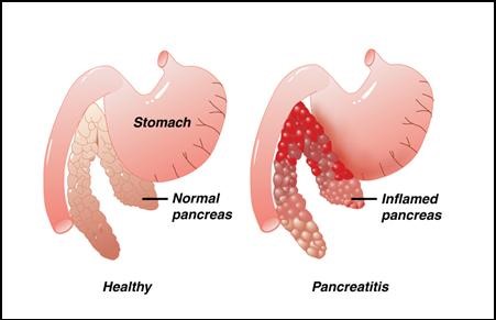
The pancreas is in the upper tummy (abdomen) and lies behind the stomach and guts (intestines). It makes a fluid that contains chemicals (enzymes) that are needed to digest food. The enzymes are made in the pancreatic cells and are passed into tiny tubes (ducts). These ducts join together like branches of a tree to form the main pancreatic duct. This drains the enzyme-rich fluid into the part of the gut just after the stomach (the duodenum). The enzymes are in an inactive form in the pancreas (otherwise they would digest the pancreas). They are ‘activated’ in the duodenum to digest food.
Groups of special cells called ‘Islets of Langerhans’ are scattered throughout the pancreas. These cells make the hormones insulin and glucagon. The hormones are passed (secreted) directly into the bloodstream to control the blood sugar level.
The bile duct carries bile from the liver and gallbladder. This joins the pancreatic duct just before it opens into the duodenum. Bile passes into the duodenum and helps to digest food.

Prevalence
Figures suggest there has been a dramatic increase in prevalence over time. The worldwide prevalence is estimated as 4-5%. Its more common in men compared to women (4:1).
Process of developing chronic pancreatitis
Although the underlying mechanism of chronic pancreatitis is murky, there have been many theories:
The most common thought is that there is obstruction or reduction of bicarbonate excretion. This in turn leads to activation of pancreatic enzymes, which leads to pancreatic tissue necrosis with eventual fibrosis. Abnormalities of bicarbonate excretion can be the result of functional defects at the level of the cellular wall, as in cystic fibrosis, or mechanical, such as trauma.
Alcohol causes proteins to precipitate in the ductular structure of the pancreas which leads to local pancreatic dilatation and fibrosis. However, this is not seen in patients with recurrent attacks of acute pancreatitis associated with alcohol use.
There may be direct toxic effects of alcohol on the pancreas. However, it has been noted that patients who drink ‘normal’ amounts of alcohol can also develop chronic pancreatitis, ie a dose-response relationship does not exist. It may be that there is excessive free radical formation which leads to pancreatic damage.
Whatever the cause, the end result is pancreatic fibrosis which can take several years to develop.
Classification of Chronic Pancreatitis
Two pathological subtypes of chronic pancreatitis: large duct pancreatitis and small duct pancreatitis are believed to be present. These two entities may represent two ends of the same spectrum but they have different pathological and radiological features and the management varies. They are usually applied to all causes of chronic pancreatitis.
Large duct pancreatitis is characterised by dilatation and dysfunction of the large ducts and is easily seen on imaging. It tends to occur more in men and there is also diffuse pancreatic calcification. Steatorrhoea is common and replacement of pancreatic enzymes does not reduce pain and patients usually require surgery to alleviate symptoms.
On the other hand, small duct pancreatitis occurs more in women and is not associated with pancreatic calcification. Imaging is usually normal, making it difficult to diagnose and, therefore, a high index of suspicion is needed. Steatorrhoea is a rare feature and patients respond to replacement of pancreatic enzymes.
Different types of chronic pancreatitis according to causes
Genetic pancreatitis:
Rare and similar to chronic pancreatitis. It presents at a younger age with epigastric pain. Most cases are thought to result from genetic mutation-induced changes in human cationic trypsinogen (PRSS1) which lead to increased auto-activation by altering chymotrypsin C (CTRC)-dependent trypsinogen activation and degradation.
Exocrine and endocrine dysfunction are also present and there is a high incidence of pancreatic carcinoma (10% at age of 50, 18.75% at age of 60, 53.5% at age of 75)
Tropical prolonged pancreatitis:
This was first described in the 1960s. It is seen in developing countries with a tropical climate and affects patients at a younger age. It has similar presentation to alcoholic chronic pancreatitis.
It is associated with large intraductal calculi and development of insulin-requiring diabetes mellitus, although ketosis is rare. Once diabetes mellitus has developed, it is often called fibrocalculous pancreatic diabetes.
One study reported a prevalence of 0.36% (1:276) of all self-reported diabetes and 0.019% (1:5,200) of the general population in Chennai in South India.
The aetiology of tropical pancreatitis is not clearly understood but may relate to malnourishment and dietary toxins – eg, in cassava.
Tropical pancreatitis is associated with SPINK1 and/or CFTR gene mutations in approximately 50% of patients.
There is an increased risk of developing pancreatic carcinoma .
Autoimmune chronic pancreatitis:
This is chronic pancreatic inflammation which results from an autoimmune process. There is a high prevalence in Japan.
It is recognised that there are two types of the condition. Type 1 is more common in Eastern Countries whilst type 2 is more common in the Western world. There are significant histological differences between the two types.
The presentation is very similar to other forms of chronic pancreatitis. There are elevated levels of serum gammaglobulins and immunoglobulin G (IgG) levels. Autoantibodies may also be increased – eg, rheumatoid factor, antinuclear antibody
Autoimmune chronic pancreatitis is steroid-responsive and reversible. There may be an association with pancreatic carcinoma.
Indoctrinate elements
One study reported that in the Western world 60-70% of patients with chronic pancreatitis have a 6- to 12-year history of alcohol abuse.
Smoking is an independent risk factor.
There has also been research into the role of genetic abnormalities in chronic pancreatitis. Genetic variation in the gamma-glutamyltransferase 1 (GGT1), CFTR and SPINK1 genes is associated with chronic pancreatitis. Chronic pancreatitis is emerging as a multifactorial condition which is the result of a complex interplay between genetically induced metabolic changes at the level of the pancreatic cell.
More aspects
There are many other causes and they are similar to those for acute pancreatitis: Iatrogenic – eg, Endoscopic retrograde cholangiopancreatography(ERCP)
Biliary tract disease
Metabolic disorders – eg, hypertriglyceridaemia, hypercalcaemia
Trauma
Congenital disorders – eg, cystic fibrosis
Abdominal radiotherapy
Drugs – eg, azathioprine, sulfonamides, loop diuretics
Idiopathic – thought to be early- and late-onset subgroups
Appearance
Appearance of chronic pancreatitis may be noticed with episodes of exacerbation with intervening remission or continuous pain in patients.
Abdominal pain. Classically, epigastric pain radiating into the back. The pathogenesis of this pain is not understood but pancreatic duct obstruction from one of the complications of chronic pancreatitis may be one cause. Usually the pain is very severe, which often requires opiates in the acute setting. In some cases pain may not be the major feature.
Decreased appetite.
Exocrine dysfunction: Malabsorption with weight loss, diarrhoea, steatorrhoea (pale, loose, offensive stools that are difficult to flush) and protein deficiency.
Endocrine dysfunction: Diabetes mellitus and its associated morbidity and mortality.
Examination can be largely unremarkable apart from tenderness in the abdomen.
Other causes with similar presentation
Acute hepatitis
Acute cholecystitis
Peptic ulcer disease
Abdominal Aortic aneurysm
Acute pancreatitis
Pneumonia
Other causes of pancreatic calcification on abdominal imaging
Severe protein malnutrition
Hereditary pancreatitis
Hyper-parathyroidism
Principles of management of chronic pancreatitis
Pain and malabsorption need to be managed.
Patients may also need assessment and counselling regarding alcohol intake and illicit drug use.
Look for the presence of psychiatric conditions – eg, depression.
History – symptoms of abdominal pain, diarrhoea, weight loss. Look for red flags such as bowel change which may indicate neoplastic disease.
Alcohol intake, smoking status, family history.
Examination – mostly to rule out other causes of abdominal pain – eg, cholecystitis.
Investigations – abdominal ultrasound, baseline blood tests including liver function to look for biliary tract obstruction, refer on for possible secretin test and ERCP or CT s
It is important to make sure that there is not another cause for the pain – eg, pseudo-cyst, duct obstruction or dysmotility.
if there is severe pain, the patient may need to be referred urgently for assessment. If there is mild-to-moderate pain, it needs to be considered whether the patient can manage at home with simple analgesia, and then refer as an outpatient to gastroenterologists/Gastro-intestinal or HPB Surgeons.
Pro-activity
Lifestyle advice regarding alcohol intake, smoking cessation, dietary advice – high in protein and low in carbohydrate. Advise abstinence from alcohol.
Pain relief
Pain relief commonly requires opiates and there is a risk of opiate dependency. Simple analgesics should be used initially – eg, paracetamol and non-steroidal anti-inflammatory drugs (NSAIDs), provided there are no contra-indications.
Opiates may also lead to gastroparesis.
ERCP may help to reduce pain by dilating strictures of the pancreatic ducts. Stenting is appropriate for single benign strictures but the stent may require changing every three months. The stricture often recurs if the stent is removed completely.
Coeliac plexus block via a gastric approach under EUS guidance has a technically high success rate with low complications. Unfortunately, its effects wear off eventually (only 10% of patients experience long-term pain relief after 24 weeks) so it is reserved as a temporary measure or where no other option is available.
Some of these procedures may result in acute pancreatitis and other complications.
Patients who remain in pain despite the above measures may need to be referred for a surgical opinion (see ‘Surgical management’, below) and if there is likely to be a delay or they are unsuitable for surgery, they should be referred to local pain clinics.
Malabsorption
Malabsorption is treated by replacing pancreatic enzymes. Diarrhoea can improve but does not usually resolve completely. Lipase is the treatment of choice but it is degraded in the stomach so that high doses have to be given.
Pancreatic enzymes: Replacement of pancreatic enzymes can help malabsorption and also reduce pain – eg, Creon. It is thought that the pancreatic enzymes provide negative feedback on pancreatic exocrine function.
Cholecystokinin (CCK) also stimulates the pancreas and is released from the small intestine by CCK-releasing peptide. The CCK-releasing peptide is broken down by proteases. Pancreatic enzymes that reach the small intestine can break down the CCK-releasing peptide, thereby halting pancreatic stimulation.
Pancreatic enzymes are administered orally and are degraded by stomach fluid reducing the bioavailability. Acid-suppressing medications can be administered at the same time with varying results.
Pancreatic enzymes are usually given as a trial for one month and, if they are effective, they are continued for six months and then they are withdrawn. 50% of patients will have long-term resolution of pain. However, if patients fail to respond then they should be considered for surgical procedures.
Octreotide
Octreotide is a somatostatin analogue and inhibits pancreatic enzyme secretion and CCK levels. It has been used with varying success rates – but this is limited by the subcutaneous route of administration.
Surgical options
Laparoscopic pancreatitis Treatment/Pancreatitis treatment may be required for the management of complications – eg, pseudo-cyst decompression. An endoscopic approach under ultrasound guidance is increasingly being used.
Pancreatic duct stones may pass spontaneously, may dislodge with ERCP or may require endoscopic shock wave lithotripsy or laser lithotripsy.
Laparoscopic pancreatitis Treatment/Pancreatitis treatment decompression of duct dilatation can be performed if this cannot be achieved by ERCP alone. Furthermore, if duct dilatation is large (eg, more than 6 mm), a lateral pancreatico-jejunostomy can be performed. Surgical drainage has proved superior to endoscopic techniques for patients with dilated ducts and chronic severe pain.
Laparoscopic pancreatitis Treatment/Pancreatitis treatment resection may also relieve pain in selected cases.
Most children and young adults with hereditary pancreatitis can be managed with endoscopic therapy but more invasive surgery is sometimes required.
Pancreatic surgical options
Pancreatoduodenectomy has been used in chronic pancreatitis and pancreatic cancer. Other forms of surgery used include Beger’s procedure (duodenal preserving resection of the pancreatic head) and Frey’s procedure (extended lateral pancreaticojejunostomy).
Radical surgery is used for relief of intractable pain not responding to other methods. This procedure has been shown to be effective in providing long-term pain relief in patients.
Recurrence of pain can occur and is often related to alcohol use. However, patients will require pancreatic enzyme supplementation, as they lose a large majority of exocrine function.
A meta-analysis reported that pancreaticoduodenectomy and duodenum-preserving pancreatichead resection were equally effective with regard to pain relief and preservation of pancreatic function. However, at long-term follow-up, duodenum-preserving procedures resulted in a better quality of life and improved long-term outcome in several measures including weight gain, diarrhoea and fatigue. Combining surgery with pancreatic enzyme replacement not only provides pain relief but is also associated with weight gain.
Latest management approaches
Pancreatectomy, followed by autologous islet cell transplantation, has been used in chronic pancreatitis. As islet cells are replaced when there is no, or less, endocrine dysfunction. The autologous cells are infused into the portal vein. It is a lengthy operation and is associated with major complications. However, it is associated with reduced pain scores and a reduction in insulin requirements.
Intricacies
Cobalamin deficiency.
Diabetes mellitus – usually brittle and associated with complications.
Pericardial/pleural/peritoneal effusions
Pseudo-cyst – localised collections of pancreatic fluid which require drainage and bowel rest.
Gastrointestinal tract haemorrhage – eg, from a pseudo-cyst invading the duodenum.
Pancreatic carcinoma – it is postulated that chronic inflammation leads to an adenocarcinoma change in pancreatic tissue.
CONCLUSION
The result of chronic inflammation of the pancreas which results in irreversible damage is chronic pancreatitis. It appears in the form of severe abdominal pain and endocrine or exocrine dysfunction. It is a difficult illness to diagnose and manage, making it a challenge for all involved.
Whether acute and chronic pancreatitis are related, is unclear although the damage to the pancreas in acute pancreatitis is reversible. At present there is no method to detect early chronic pancreatitis and calcification of the pancreas may not develop until 5-10 years after initial symptoms.
Chronic pancreatitis is associated with increased mortality and morbidity – one third of patients will die within 10 years. Furthermore, for those who continue to drink alcohol, this risk is further increased. The exception is autoimmune pancreatitis which responds well to steroids, even if relapses occur.
There is increased incidence of pancreatic cancer in patients with chronic pancreatitis though the association is not proven beyond doubt. Or there is no concrete evidence to prove the association.